A Chromatin Landmark and Transcription Initiation at
Most Promoters in Human Cells
Data
Figures
Supplemental Information
Acknowledgements
References
|
Figure 7

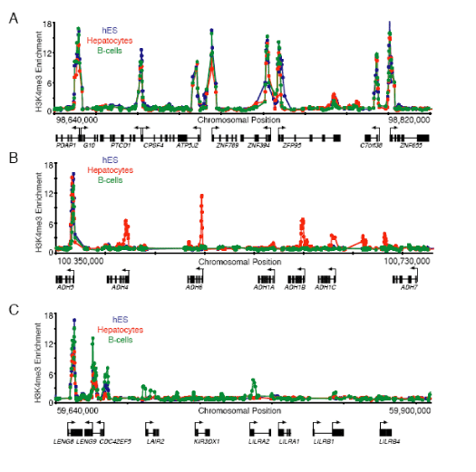
Figure 7. Tissue specific methylation of H3K4 at gene clusters
A. Enrichment ratios for H3K4me3 from hES cells (blue), primary human hepatocytes (red) and the REH line of pro-B cells (green) across 180kb of chromosome 7 are shown. Start sites and direction of transcription are indicated by arrows.
B. Enrichment ratios for H3K4me3 from hES cells (blue), primary human hepatocytes (red) and the REH line of pro-B cells (green) across 380kb of chromosome 4 spanning the alcohol dehydrogenase cluster are shown. ADH1A is specifically expressed in fetal liver and ADH7 is expressed in gastric tissue. Constitutively H3K4me3-modified ADH5 is expressed in all samples tested.
C. Enrichment ratios for H3K4me3 from hES cells (blue), primary human hepatocytes (red) and the REH line of pro-B cells (green) across 240kb of chromosome 19 spanning the leukocyte receptor cluster are shown. Start sites and direction of transcription are indicated by arrows.
|

