Figure S3
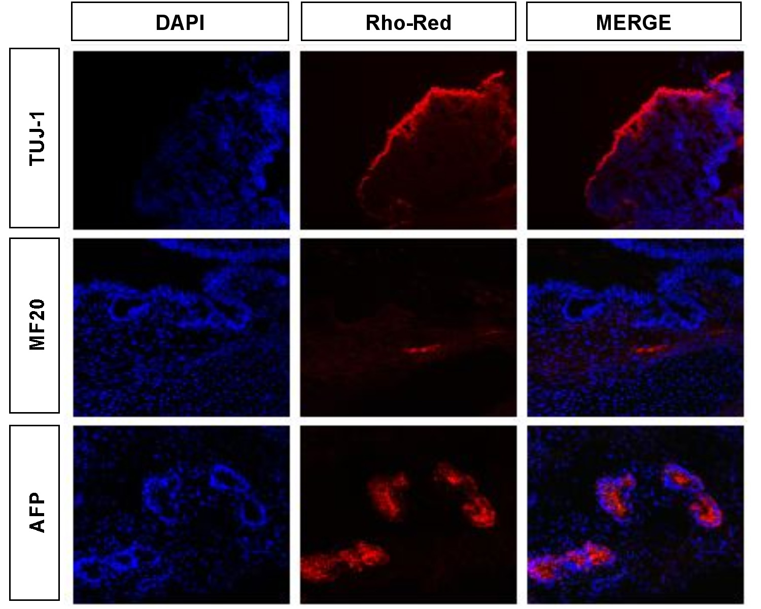
Figure S3. Analysis of human ES cells for differentiation potential. Teratomas were analyzed for the presence of markers for ectoderm (Tuj1), mesoderm (MF20) and endoderm (AFP). For reference, nuclei are stained with DAPI. Antibody reactivity was detected for derivatives of all three germ layers confirming that the human embryonic cells used in our analysis have maintained differentiation potential.
|